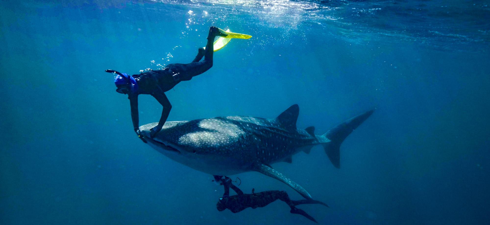
An international team of researchers has discovered a new method of imaging free-swimming whale sharks using underwater ultrasound.
The research, published in Frontiers, was led by the Australian Institute of Marine Science (AIMS) in a collaboration with The University of Western Australia, WA’s Mira Mar Veterinary Hospital, Okinawa Churaumi Aquarium in Japan, and Georgia Aquarium in the US.
Lead author Dr Mark Meekan, from UWA’s Oceans Institute, has been running a monitoring program at Ningaloo Reef with AIMS for the past 20 years.
“Whale sharks are large filter feeders which makes them vulnerable to consuming plastics and man-made chemicals in the water, so we want to know if they’re healthy,” Dr Meekan said.
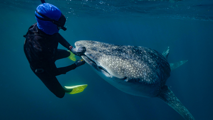
As part of the program the researchers have been collecting tiny parasites called copepods, a small shrimp-like animal, from the whale sharks’ lips and edges of their fins.
“We found when we started to scrape the copepods off their lips, the whale sharks slowed down, hung vertically in the water and treated us like a giant cleaner fish,” Dr Meekan said.
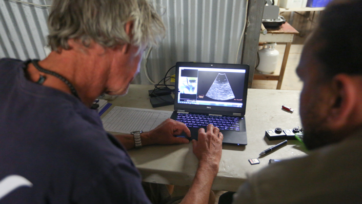
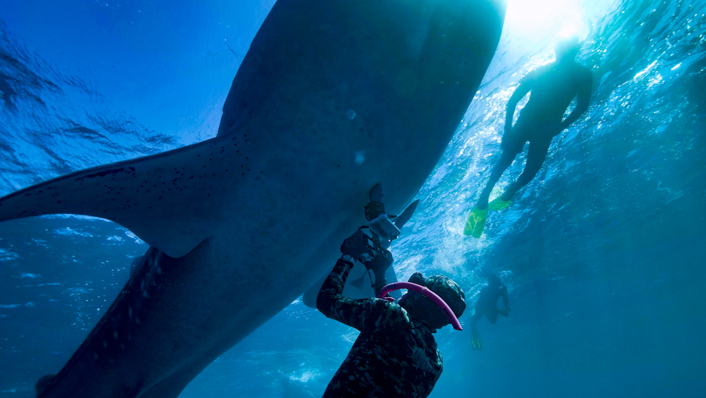
While the whale sharks were in this position the researchers were able to use an underwater ultrasound to capture images of the internal organs to help to assess their condition and reproductive status.
“Underwater ultrasounds have been used before to look at reproductive status of sharks in aquariums or caught on drumlines, but this method is not viable with whale sharks,” Dr Meekan said.
Kim Brooks was AIMS’ senior field technician during the expedition and operated the hand-held ultrasound unit while free diving with a dozen whale sharks.
“We began by trying to find landmarks inside the body, starting with the heart and from there tried to work out where we were in relation to the rest of the organs,” Mr Brooks said.
“It was both an awesome and challenging experience because whale sharks are the largest fish in the ocean, and I was able to watch a live screen view of their beating heart while holding my breath underwater.”
Dr Meekan said the ultrasound imaging showed that whale sharks have a very slow heart rate – just 12 to 16 beats per minute.
The researchers then started to map the internal organs and at first were interested in the liver, where oil is stored to keep the whale shark buoyant.
“We also imaged the back of the shark and could clearly see the skin thickness and muscle bundles,” Dr Meekan said.
“They have a layer of hard denticles at the skin surface that feels rough like sandpaper and below that connective tissue up to 20cm deep, making their skin one of the thickest of any animal.
“We found whale sharks that were skinny and in poor condition had thinner skins.”
Scuba diving around whale sharks is not permitted at Ningaloo and touching whale sharks is illegal.
The research was conducted and funded by AIMS and supported by Santos. It had ethics and sampling permits granted by management authority the WA Department of Biodiversity, Conservation and Attractions before any work was attempted.


